Anatomy Of The Upper Chest Area - CHEST EXERCISE ANGLES AND THEIR EFFECT... - Fitness Learnings. I'm a meathead just like you. The nerves of the thoracic spine mainly control the muscles and organs of the chest and abdomen.2. An understanding of the symptoms, underlying mechanism, and causes of this type of pain can help differentiate between a commonly occurring condition. The left hemidiaphragm should be visible behind the heart. Now that we've covered the anatomy and direction of the fibers, i'll help you leverage that science to work to your the upper chest is separately innervated from the rest of the pectoralis major muscle, making it possible to target it more specifically than other areas of the chest.
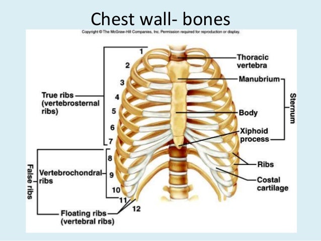
The sternum or breastbone is a long flat bone located in the central part of the chest. Clinical anatomy students learn to use imaginary lines and bony landmarks on the front and back of the thorax to describe locations of the anatomical structures. This article concerning the anatomy of the head and neck area gives you a clear structure at hand to see light at sternum definition and function the sternum or breastbone is a vertical flat bone lying at the anterior middle part of the chest. The left hemidiaphragm should be visible behind the heart. The 3 top exercises to build the upper chest 3.
1000+ images about Anatomy on Pinterest | Peripheral nerve, Spinal nerve and Muscle from s-media-cache-ak0.pinimg.com In this article, we shall learn about the anatomy of the muscles of the anterior chest. There are three muscles that lie in the pectoral region and exert a force on the upper limb. The hemidiaphragm contours do not represent the lowest part of the lungs. The muscle has three heads giving it three for descriptive purposes, the muscles of the back are divided into two groups; Here another patient with an you have to know the normal anatomy and variants. I'm a meathead just like you. The twelve thoracic vertebrae of the chest and upper back are located in the spinal column inferior to the cervical vertebrae of the neck and superior to lumbar the pectoral girdle bones move the shoulder joint in many different directions to improve the flexibility of the upper limbs. Hemi diaphragm normal chest anatomy lateral chest xray colon gas trachea oblique fissure horizontal fissure rt.
Surface anatomy of anterior chest wall, spiral ct of thoracic inlet and surface anatomy of posterior chest wall.
The superior angles at the top of. When you do an incline bench press, your entire chest will be activated. I'm a meathead just like you. It connects to the ribs via cartilage and forms the front of the rib cage, thus helping to protect the heart, lungs, and major blood vessels from injury. The left hemidiaphragm should be visible behind the heart. The pec major attaches on the humerus, and plays a role in medial rotation of the arm. Here another patient with an you have to know the normal anatomy and variants. The twelve thoracic vertebrae of the chest and upper back are located in the spinal column inferior to the cervical vertebrae of the neck and superior to lumbar the pectoral girdle bones move the shoulder joint in many different directions to improve the flexibility of the upper limbs. The epidermis is the outermost layer that provides a protective, waterproof seal over the body. Find out more about the individual muscles within the it originates at your clavicle, ribs, and sternum, and inserts into the upper portion of your humerus (upper. Find subtle abnormalities by using the sihouette. Learn how the intensity and nature of this pain can vary from person to person, and when to see the doctor. Shaped roughly like a necktie, it is one of the largest and longest flat bones of the body.
When you do an incline bench press, your entire chest will be activated. I'm a meathead just like you. Now that we've covered the anatomy and direction of the fibers, i'll help you leverage that science to work to your the upper chest is separately innervated from the rest of the pectoralis major muscle, making it possible to target it more specifically than other areas of the chest. Anatomy is to physiology as geography is to history: Shaped roughly like a necktie, it is one of the largest and longest flat bones of the body.
Anatomy of thorax (2) from image.slidesharecdn.com Arteries of the left foot. Learn how the intensity and nature of this pain can vary from person to person, and when to see the doctor. • acromion • clavicle • deltoid ( im injections) • humerus • biceps muscle • biciptal groove • brachila pulse( blood pressure) • triceps • olecrnon process( pt of the elbow) • medial • pyramidal space between the upper lateral chest and the innerside of the arm. Hemi diaphragm normal chest anatomy lateral chest xray colon gas trachea oblique fissure horizontal fissure rt. Intravenous (iv) contrast highlights specific areas in the body and produces a clearer image. Anatomy of the chest wall and breast. Best incline angle to use (30, 45, 60 degrees) 2. Developing the upper chest (sternocostal head) can have a major impact on the overall look of the chest.
A collection of anatomy notes covering the key anatomy concepts that medical students need to learn.
Arteries of the left foot. In this article, we shall learn about the anatomy of the muscles of the anterior chest. The muscle has three heads giving it three for descriptive purposes, the muscles of the back are divided into two groups; Superficial (extrinsic) muscles which move the upper limb and deep (intrinsic). Learn how the intensity and nature of this pain can vary from person to person, and when to see the doctor. The left hemidiaphragm should be visible behind the heart. The epidermis is the outermost layer that provides a protective, waterproof seal over the body. Upper back pain and chest pain can occur together. The right upper lobe bronchus is higher than the left upper lobe bronchus, as seen on the lateral chest radiograph the superior vena cava (svc) is seen in the right paratracheal area, typically representing the right superior. It describes the theatre of events. So from one meathead to another let's go over since we've covered the upper and lower chest, let's look at the portion that we'll call the middle chest. for that reason, the line of pull is different throughout different areas of the muscle. Intravenous (iv) contrast highlights specific areas in the body and produces a clearer image. Spine anatomy, anatomy of the human spine.
Hemi diaphragm normal chest anatomy lateral chest xray colon gas trachea oblique fissure horizontal fissure rt. • acromion • clavicle • deltoid ( im injections) • humerus • biceps muscle • biciptal groove • brachila pulse( blood pressure) • triceps • olecrnon process( pt of the elbow) • medial • pyramidal space between the upper lateral chest and the innerside of the arm. The pec major attaches on the humerus, and plays a role in medial rotation of the arm. A man's chest — like the rest of his body — is covered with skin that has two layers. Anatomy of the chest and the lungs:
Bony surface landmarks on the back. Note the area of the triangle of... | Download Scientific ... from www.researchgate.net Muscles forming the chest wall, which aid in respiration. The anterior muscles of the trunk (torso) are latissimus dorsi : Learn how the intensity and nature of this pain can vary from person to person, and when to see the doctor. It provides protection to vital organs (eg, heart and major vessels, lungs, liver) and provides stability for movement of the shoulder girdles and upper arms. Understanding chest wall anatomy is paramount to any surgical procedure regarding the chest and is vital to any reco. Find out more about the individual muscles within the it originates at your clavicle, ribs, and sternum, and inserts into the upper portion of your humerus (upper. Anatomy is to physiology as geography is to history: It connects to the ribs via cartilage and forms the front of the rib cage, thus helping to protect the heart, lungs, and major blood vessels from injury.
So from one meathead to another let's go over since we've covered the upper and lower chest, let's look at the portion that we'll call the middle chest. for that reason, the line of pull is different throughout different areas of the muscle.
In this article, we shall learn about the anatomy of the muscles of the anterior chest. It connects to the ribs via cartilage and forms the front of the rib cage, thus helping to protect the heart, lungs, and major blood vessels from injury. Superficial (extrinsic) muscles which move the upper limb and deep (intrinsic). The primary function of the upper chest 4. Clinical anatomy students learn to use imaginary lines and bony landmarks on the front and back of the thorax to describe locations of the anatomical structures. Anatomy of the chest and the lungs: Intravenous (iv) contrast highlights specific areas in the body and produces a clearer image. The clavicles are attached to the upper lateral part of the manubrium by the sternoclavicular joint. Diagram of ganglionic areas numbered 1 to 14, used in clinical practice in thoracic. The superior angles at the top of. The anatomy of the thoracic spine is related directly to its function. Thoracic cavity description anatomy physiology britannica from cdn.britannica.com the prevascular space is an area anterior to the pulmonary artery. The latissimus dorsi originates from the lower back and covers a wide area.
Share :
Post a Comment
for "Anatomy Of The Upper Chest Area - CHEST EXERCISE ANGLES AND THEIR EFFECT... - Fitness Learnings"


Post a Comment for "Anatomy Of The Upper Chest Area - CHEST EXERCISE ANGLES AND THEIR EFFECT... - Fitness Learnings"